
Fig. 3.
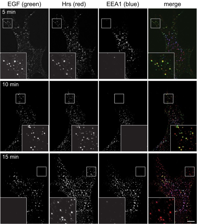
EGF passes through peripheral Hrs endosomes prior to labelling EEA1 endosomes. RPE cells were serum starved, then pulsed for 2 min with fluorescent EGF, then chased for a further 3 min (upper row), 8 min (middle row) or 13 min (lower row) before fixing and labelling with antibodies against Hrs or EEA1. The insets show a ×3 magnification of the indicated areas. Scale bar: 10 µm.