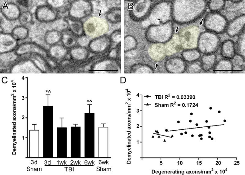
FIGURE 4.

Intact axons undergoing demyelination after TBI. (A, B) Demyelinated axons (light yellow) have normal mitochondria and cytoskeletal structure but lack a myelin sheath. Only axons with a diameter greater than 0.3 μm were counted as demyelinated to avoid potential inclusion of unmyelinated axons. (C) Demyelinated axons are significantly increased after TBI but only at 3 days and 6 weeks. * p ≤ 0.01 compared to 3-day sham. ^p ≤ 0.05 compared to 6-week sham. (D) Values for demyelinated axons do not correlate with values for degenerating axons within individual animals across the post-TBI time course (3 days to 6 weeks). Image time points: (A) 3-day TBI; (B) 6-week TBI. Scale bar = 1 μm. d, days; wk, weeks.
