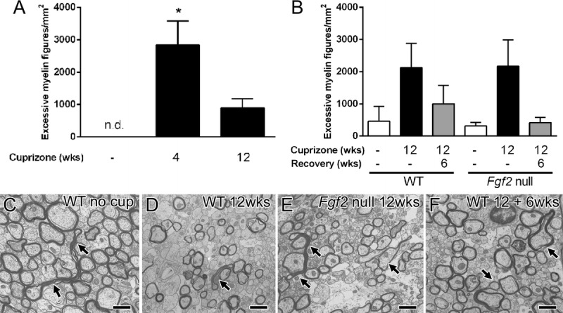
FIGURE 7.

Excessive myelin figures during cuprizone-induced demyelination and remyelination. (A) In C57BL/6 mice, cuprizone ingestion produces widespread demyelination of the corpus callosum. Cuprizone treatment for 4 weeks results in oligodendrocyte loss, initial demyelination, and axon damage. Prolonged cuprizone administration through 12 weeks produces chronic demyelination. Excessive myelin figures are not detected (n.d.) in 8-week-old mice not taking cuprizone but are significantly increased during the period of initial demyelination (* p = 0.0009). (B–F) Excessive myelin figures were also analyzed relative to the extent of remyelination. Removal of cuprizone from the diet allows for endogenous progenitor cells to attempt to generate new oligodendrocytes and to initiate remyelination. Remyelination is more extensive in Fgf2-null mice in comparison with wild-type (WT) mice (29). Excessive myelin figures are similar between WT and Fgf2-null mice without cuprizone (26 weeks of age controls), during chronic demyelination (12 weeks), and after a subsequent 6-week period on a normal diet to allow for remyelination (12 + 6 weeks). (C–F) Arrows indicate examples of excessive myelin figures present in all genotypes and time points. Scale bar = 1 μm. wks, weeks.
