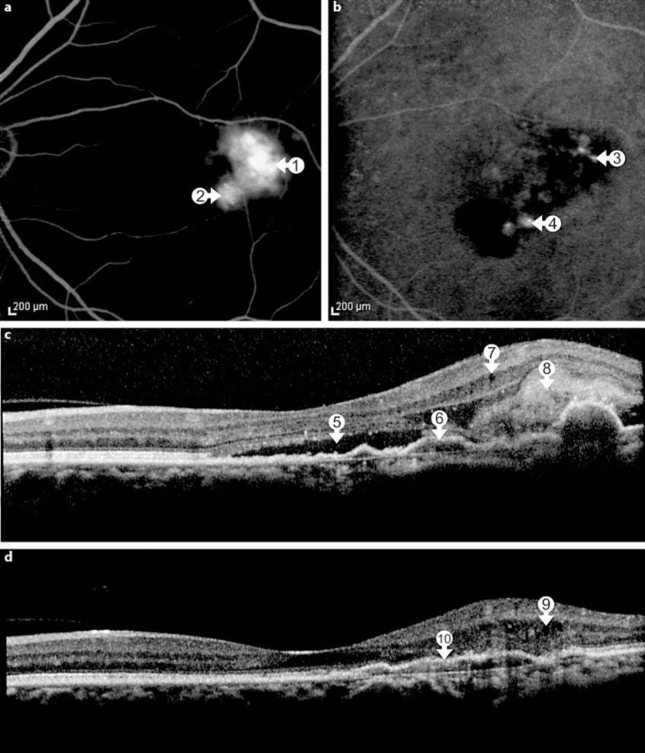
Fig. 2.
a Two years later, FA identifies two active leakage sites temporal and inferior to the fovea (arrows 1 and 2). b Indocyanine green angiography reveals 2 separate hot spots, temporal and inferior to the fovea (arrows 3 and 4). c SD-OCT detects subretinal fluid (arrow 5) and an enlarged area with RPE irregularities and RPE detachment (arrow 6), intraretinal cysts (arrow 7) and hyperreflective subretinal material (arrow 8). d SD-OCT following successful treatment with ranibizumab injections shows persistent small intraretinal cysts (arrow 9) and a fibrovascular RPE detachment temporal to the fovea (arrow 10).

