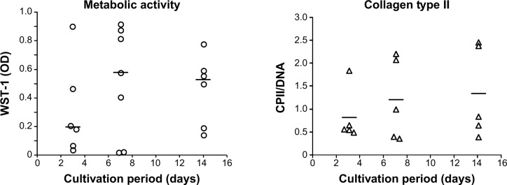
Figure 3.

Metabolic cell activity and matrix production of primary chondrocytes cultured on the collagen-based scaffold.
Notes: Chondrocytes were isolated from the hyaline cartilage of patients (n≥5) undergoing primary total knee joint replacement and seeded (1×10⁵ cells/cm2) onto the scaffold before being incubated for 3, 7, and 14 days. The metabolic activity was measured via WST-1 (left panel). The production of CPII was quantified and normalized to the DNA content per patch (right panel). Each symbol (circle, triangle) represents a donor. Black lines correspond to the median. Analysis of variance post hoc-LSD was conducted (no significant differences between 3, 7, and 14 days of cultivation).
Abbreviations: CPII, procollagen type II; LSD, least significant difference; OD, optical density; WST-1, water-soluble-tetrazolium salt.
