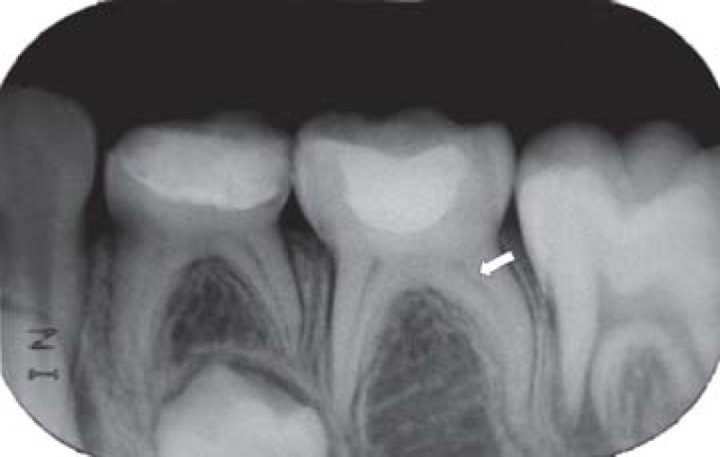
FIGURE 2B. 3-month follow-up periapical radiograph suggesting the initial formation of a dentin bridge immediately below the Portland cement in the distal root (arrow) of the pulpotomized mandibular left second molar and absence of periapical lesion in both pulpotomized mandibular left primary molars.

