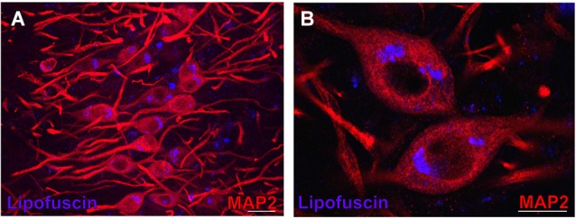
Figure 3.

Lipofuscin granules in medial superior olive (MSO) neurons of an aged gerbil. MSO neurons are MAP2 immunostained (Alexa Fluor 647, red). Lipofuscin granules have been excited with the DAPI excitation wavelengths and appear blue. Confocal images show a maximum projection of image stacks in (A) and a single optical image of 0.3 µm thickness in the enlargement in (B). Scale bar in (A): 50 µm and 20 µm in (B).
