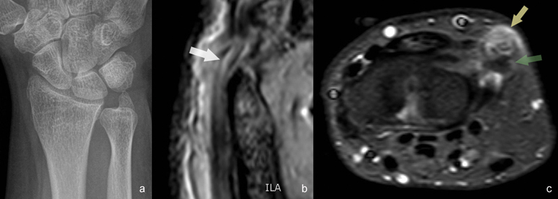
Fig. 1a–c.

(a) Anteroposterior (AP) radiograph showing oversized ulnar styloid. (b) T2-weighted sagittal MRI image showing internal longitudinal lesions (white arrow) where the ECU tendon hooks around the ulnar styloid. (c) STIR-weighted axial MRI image showing important tenosynovitis (yellow arrow) at the tip of the ulnar styloid (green arrow).
