
Fig. 3a, b.
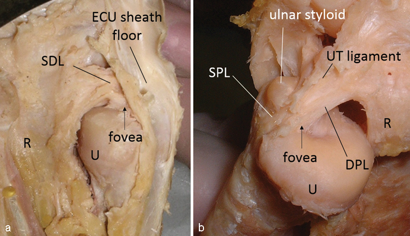
Ulnar insertions of the TFCC on a cadaver specimen (right wrist). (a) TFCC seen from the dorsal side of the ulna. The SDL and the floor of the extensor carpi ulnaris (ECU) tendon sheath hinder the view of the fovea. (b) The foveal insertion seen from the palmar side of the ulna. Note that TFCC insertions to the fovea and the ulnar styloid are clearly visualized. R: radius, U: ulna.