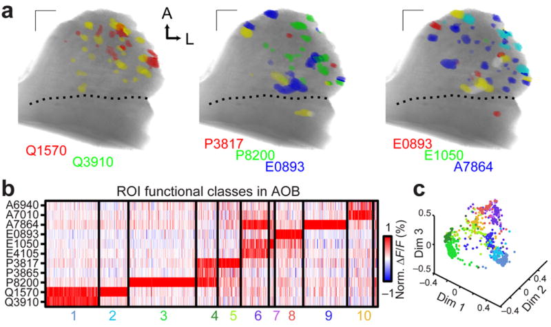
Figure 6. Glomerular activity patterns identify functional VSN classes.

(a) Glomeruli responding to the indicated steroids (10 μM) with ΔF/F > 1% were colorized according to stimulus. Glomeruli responding to more than one steroid are indicated by color mixtures (yellow: red/green, magenta: red/blue, cyan: green/blue, white: red/green/blue). Scale bars: 100 μm. (b) 1078 glomerular responses across 10 experiments to the 11-steroid panel match previously-identified VSN functional classes. Each thin vertical stripe shows the activity pattern of a single glomerular ROI, and each row shows responses to a single sulfated steroid (10 μM). A small unclustered group (far right) consisted of less than 1% of all ROIs. (c) Multidimensional scaling (first 3 dimensions) of the ROI responses to the 11 sulfated steroids in the stimulus panel. Colors of the points correspond to the clusters in b.
