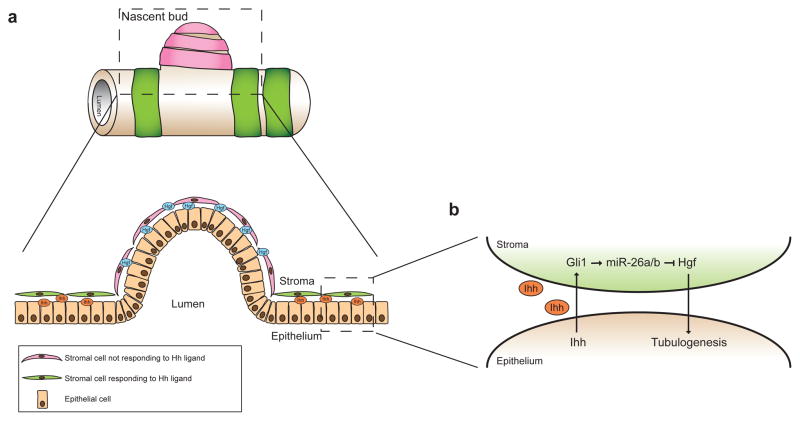
Figure 8. Model of the spatial regulation of branching by Hh and Hgf signaling.
(a) Cartoon of a section of a prostate tubule with a nascent bud. Green bands represent Hh-responsive stromal cells and pink bands represent stromal cells that are not responding to the Hh ligand. A lateral-section through the boxed region is shown below. (b) Enlarged view of the epithelial-stromal interaction in the boxed region. In regions that are not undergoing branching, Ihh secreted by epithelial cells induces expression of Gli1 in the adjacent stromal cells, which increases expression of miR-26a and miR-26b, which inhibit Hgf production, thereby preventing tubulogenesis. In regions lacking Ihh expression, surrounding stromal cells do not respond to the Hh ligand, hence Hgf levels are high and branching is induced.

