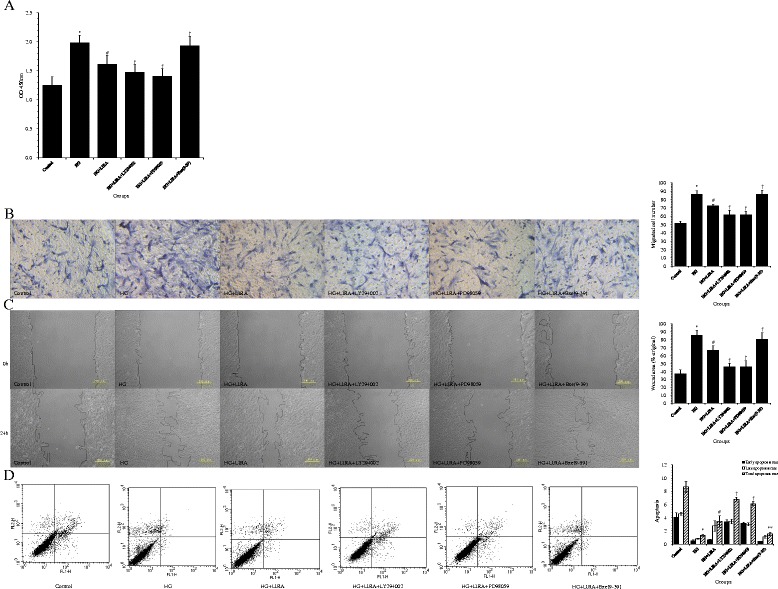
Figure 7.

Liraglutide (LIRA) exerted beneficial effects on cultured vascular smooth muscle cell (VSMCs) by activating the GLP-1 receptor and inhibiting PI3K/Akt and ERK1/2 signaling pathways. (A) Cell proliferation of VSMCs; * p < 0.01 vs. control; # p < 0.01 vs. HG; † p < 0.05 vs. HG + LIRA. (B) Representative photomicrographs showing migration of VSMCs in the Transwell migration assay (200× magnification). The relative amounts of migrating cells in all groups are presented; * p < 0.01 vs. control; # p < 0.01 vs. HG; † p < 0.01 vs. HG + LIRA. (C) Representative photomicrographs show the migrating cells in the scratch wound assay; * p < 0.01 vs. control; # p < 0.01 vs. HG; † p < 0.05 vs. HG + LIRA. (D) Apoptosis rates were determined by flow cytometry, with the lower right and upper right quadrants representing early apoptotic and late apoptotic cells, respectively. These values were quantified, and the total apoptotic rate is the sum of the early and late apoptotic rates; * p < 0.01 vs. control; # p < 0.05 vs. HG; † p < 0.01, ** p < 0.05 vs. HG + LIRA. Data from three independent experiments are expressed as mean ± standard deviation.
