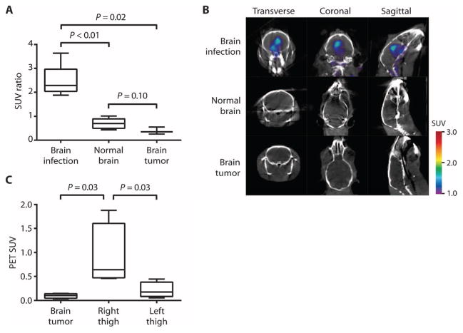
Fig. 3. 18F-FDS PET/CT imaging of human brain tumor xenograft versus E. coli infection.
(A) 18F-FDS PET signal from an E. coli–infected brain compared with a human brain tumor (U87MG) xenograft and normal brain. Data are median SUV ratios normalized to the uninflamed thigh muscle of each animal, with interquartile and ranges shown (n = 6 animals for brain infection, n = 6 for normal brain, and n = 3 for brain tumor). (B) 18F-FDS PET signal in the E. coli–infected brain, normal brain, or tumor brain tissues. (C) 18F-FDS PET signal from right-thigh E. coli infection compared with human brain tumor (U87MG) xenograft and infected left thigh in the same animals. Data are median SUVs with interquartile and ranges shown (n = 4 animals). P values in (A) and (C) were determined by two-tailed Mann-Whitney U test.

