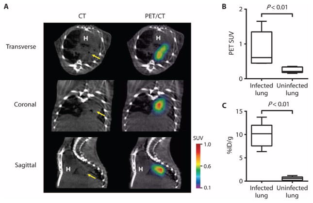
Fig. 5. K. pneumoniae lung infection (pneumonia).
(A) 18F-FDS PET signal colocalized with K. pneumoniae lung infection noted on CT (yellow arrow). (B and C) 18F-FDS PET signal in vivo (B) and postmortem bio-distribution (C) in infected versus uninfected lungs. Data are medians with interquartile and ranges shown (n = 5 animals for the infected group and n = 6 for the uninfected group).

