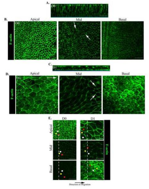
Figure 2. Alterations in cell morphology, cell positioning and organization of actin cytoskeleton accompany the transition from a stationary to a migratory epithelial phenotype.
Ex vivo wounded epithelial cultures at D0 and D1 post-injury labeled with fluorescent-conjugated phalloidin to examine cell structure, position, and F-actin organization were examined by confocal microscopy imaging. Images were collected as z-stacks at multiple planes along the epithelial cells’ apical-basal axis. Shown are (B, D, E) single optical planes at the cells’ apical, mid, and basal regions and (A, C) an orthogonal view of the collected z-stack. (E) To follow the alignment of cells within the monolayer from their apical to basal domains each cell’s position is marked by a different color dot in the apical, mid, and basal planes of the epithelium. (A, E) At D0 the cells were organized as a tightly packed cuboidal epithelium linearly aligned from their apical to basal domains. As cells entered the CMZ they dramatically altered their morphology decreasing in height (z) while increasing their spread in the x-y plane. (C, E) Cells moved at angles to one another, their basal regions typically preceding their apical domains in the direction of migration. (A,B,E) At D0 F-actin was organized as cortical actin filaments most concentrated in the cells apical domains. (C,D,E) At D1 actin remained cortical in the cells apico- and mid-lateral domains but assembled de novo lamellipodia actin filaments and stress fibers along the cells’ basal surfaces. White arrows denote F-actin concentrated at cell vertices. White long arrow indicates the direction of migration. Mag. Bar = 20 μm. Studies are representative of at least three independent experiments.

