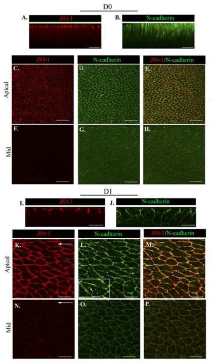
Figure 3. The apical junctional complex is maintained as cells migrate into the CMZ.
Wounded epithelial cultures were imaged at D0 and D1 after injury following immunostaining for (A,C,F,I,K, N) ZO-1 and (B,D,G,J,L,O) N-cadherin. (E,H,M,P) ZO-1/N-cadherin overlays. Z-stacks were collected and shown as (A,B,I,J) orthogonal cuts or (C-H, K-P) single optical planes at the cells’ apical and mid-lateral domains. At D0 ZO-1 is largely restricted to the cells’ apical domains (A,C). N-cadherin is similarly localized but also extends basally along the cells’ lateral borders, although at diminishing concentrations (B,D,G). At D1 there was little change in the distribution of ZO-1 (I,K) but increased levels of N-cadherin junctions in the cells’ mid-lateral zones (J,O). A higher magnification image included as an inset in (L), short white arrow indicates N-cadherin puncta. Arrows indicate the direction of migration. Mag. Bar = 20 μm. Studies are representative of at least three independent experiments.

