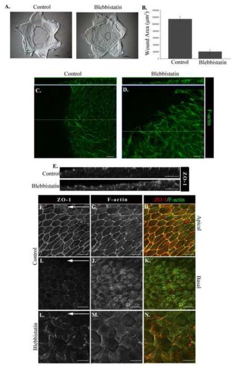
Figure 7. Myosin function is necessary for effective wound repair.
Wounded lens epithelia were cultured for one day in the presence or absence of the myosin II inhibitor blebbistatin. (A) Phase contrast imaging showed faster migration onto the wounded area in the presence of blebbistatin (Mag. Bar = 500μm), data quantified in (B). Wounded cultures were examined by confocal imaging following labeling for F-actin (C,D,G,H,J,K,M, N), or ZO-1 (E,F,H,I,K, L,N) to analyze cell shape and cell-cell connectivity. Control study in F-K was imaged at the cells’ apical (F-H) and basal (I-K) domains. Images shown are either a representative single optical plane from a z-stack (C-D, F-N) or an orthogonal cut of a collected z-stack (E). Loss of myosin function resulted in extensive disorganization of the monolayer, discontinuity of cell-cell junctions, loss of cortical actin, and disorganization of basal actin structures. Mag. Bar = 20μm. Studies are representative of at least three independent experiments.

