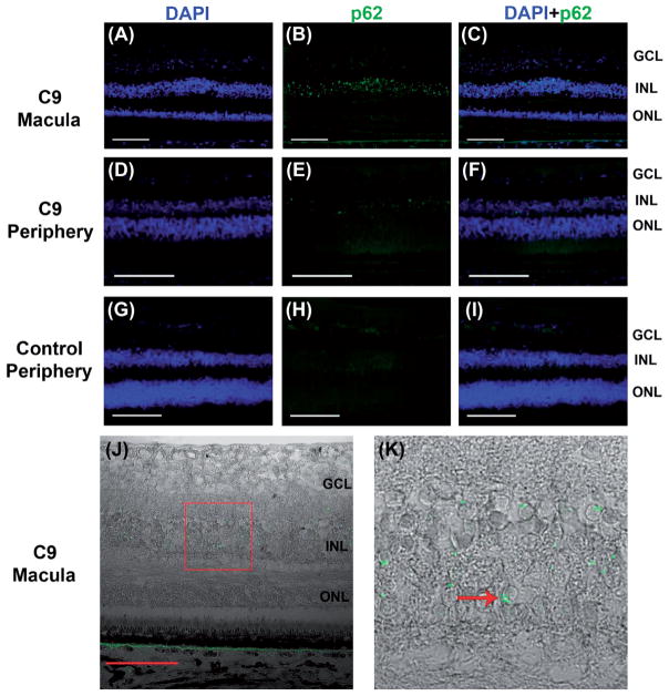
Figure 3.
p62 + immunofluorescence in C9orf72 retina and absence of inclusions in control eye. Abundant p62+ (green) inclusions are seen in the inner nuclear layer of C9-macula (A–C) and peripheral retina (D–F). No p62 + inclusions are seen in control eye (G–I). Sections are counterstained with DAPI (blue). Perinuclear location and crescent morphology of p62 + inclusions in C9orf72 macula are demonstrated on DIC imaging (J, K – inset, arrow). Scale bars = 100 μm.

