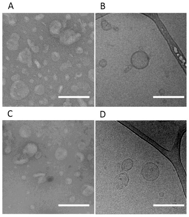
Figure 4. The nanostructures formed by L4F fusions are peptide vesicles.
TEM and Cryo-TEM were used to characterize purified L4F-A192 that was A–B) unfiltered and C–D) filtered through a 0.2 μm membrane. A, C) TEM images of negatively stained L4F-A192 revealed evidence of nanoparticles. Unfiltered, nanoparticles averaged 41 ± 16 nm and filtered vesicles are 28 ± 11 nm. B, D) Similarly, cryo-TEM revealed the presence of unilamellar vesicles with an average radius of 36 ± 13 nm unfiltered and 43 ± 17 nm when filtered. Consequently, the average membrane thickness of unfiltered particles is 8.4 ± 1.3 (n = 18) and 0.2 um filtered particles is 6.8 ± 0.7 nm (n = 8). The scale bar indicates 200 nm.

