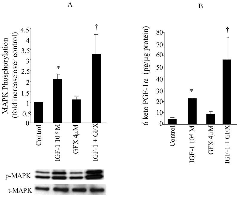
Figure 6. Role of PKC in IGF-1 induced MAPK phosphorylation and PGI2 (6-keto-PGF1α) production.
VSMC were stimulated with either BK IGF-1 (10−8M) for 10 min in the presence and absence of PKC inhibitor bisindolylmaleimide (GFX, 2μM). MAPK phosphorylation (p42mapk and p44mapk) were measured by immunoblot using anti-phosphotyrosine-MAPK antibodies (p-MAPK) and total MAPK was measured in the same immunoblot by stripping the membrane and re-immunoblotting with anti-total MAPK antibodies (t-MAPK). Release of 6-keto-PGF1α into the media, was measured by RIA. Data are expressed as mean±SE and the bar graphs are representative of 5 separate experiments. * P<0.05 vs. control, †P<0.05 vs. IGF-1.

