
Figure 3.
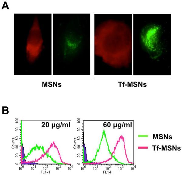
a) Fluorescence microscopy images of cell (red) and MSNs (green) show increased fluorescence of the Tf-MSNs than the untargeted MSN, indicative of higher endocytosis of Tf-MSN; and b) Flow cytometry of targeted and untargeted MSNs at two different concentrations show Tf-MSNs have a significantly higher uptake.