
Figure 4.
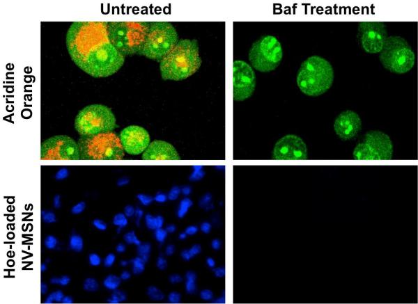
Cells (green) are treated with acridine orange to detect acidic organelles (orange). Once exposed to Bafilomycin (Baf), the organelles are no longer acidic and no orange fluorescence is seen. Hoechst-loaded NV-MSNs show nuclear staining of cells (blue) but after Baf treatment, the NVs are not activated and no staining is seen.