
Figure 5.
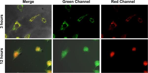
Confocal images of Dox-loaded MSNs: the green channel represents the MSNs and the red channel shows doxorubicin. At 3 hours, the Dox is contained in the MSNs located inside the cell, but at 12 hours the nanovalves have been activated and the Dox has begun to stain the cell nuclei red.