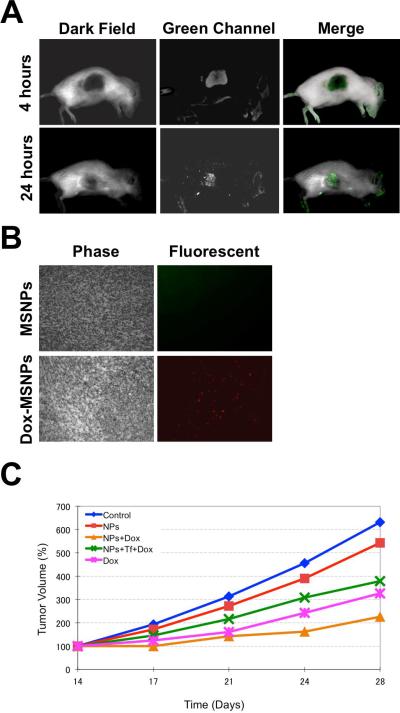
Figure 7.
a) Biodistribution of MSNs in mice with xenograft tumors. SCID mice bearing subcutaneous human pancreatic tumors were injected via tail vein with MSNs. 4 and 24 hours later, the mice were anesthetized, subjected to Maestro 2 in vivo imaging system for green fluorescent (MSNs) images. b) Tumors were collected, processed, and analyzed with fluorescence microscope. Red fluorescence indicates doxorubicin in the tumors. c) 25 SCID mice with established xenografts of MiaPaCa-2 were randomly divided to 5 groups (n=5), and the intraperitoneal injections of MSN solutions began once the average tumor diameter reached 3 mm (14th day after inoculation). All injections were done twice per week until the end of the experiment (the 28th day). The average tumor volumes are shown as means ± the standard deviation (SD).

