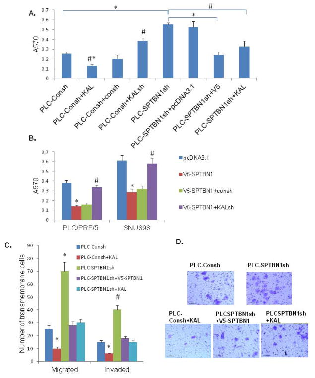
Figure 7.
Loss of SPTBN1 promotes malignant behaviors of HCC cell lines, which can be reversed by induction of Kallistatin or SPTBN1. A. As shown in PLC/PRF/5 HCC cells, pretreated with Kallistatin decreases the adhesion capacity of HCC cell (#P<0.05 when bar 2 compared with bar1 and bar3,* P<0.01 when bar 2 compared with bar 4–8); Knock-down Kallistatin increases the adhesion capacity of HCC cells (#P<0.05 when bar 4 compared with bar 3 and bar1). Knock-down SPTBN1 increases the adhesion capacity of HCC cells (*P<0.01, bar 5 compared with bar 1), that is reversed by over-expression of SPTBN1 (*P<0.01, bar 7 compared with bar 5) or pretreated with Kallistatin (#P<0.05 bar 8 compared with bar 5). B. As demonstrated in two HCC cell lines, PLC/PRF5 and SNU398, the adhesion capacity of HCC cells is inhibited by over-expression of SPTBN1 (*P<0.01, V5-SPTBN1 compared with pcDNA3.1 group), which is reversed by knocking-down Kallistatin gene (#P<0.05 when KALsh compared with Consh). C. As demonstrated in PLC/PRF/5 cells, HCC cells with decreased SPTBN1 migrate and invade two to three times as much as control cells (*P<0.01, #P<0.05 when SPTBN1sh compared with Consh), which can be reversed by over-expression of SPTBN1 or pretreated with Kallistatin (*P<0.01, #P<0.05 when SPTBN1sh compared with V5-SPTBN1 and KAL group respectively). Kallistatin inhibited malignant behaviors of PLC-Consh cells (*P<0.01 when bar2 compared to other group). D. Representative image of cells in the migration assay stained with 0.5% crystal violet.

