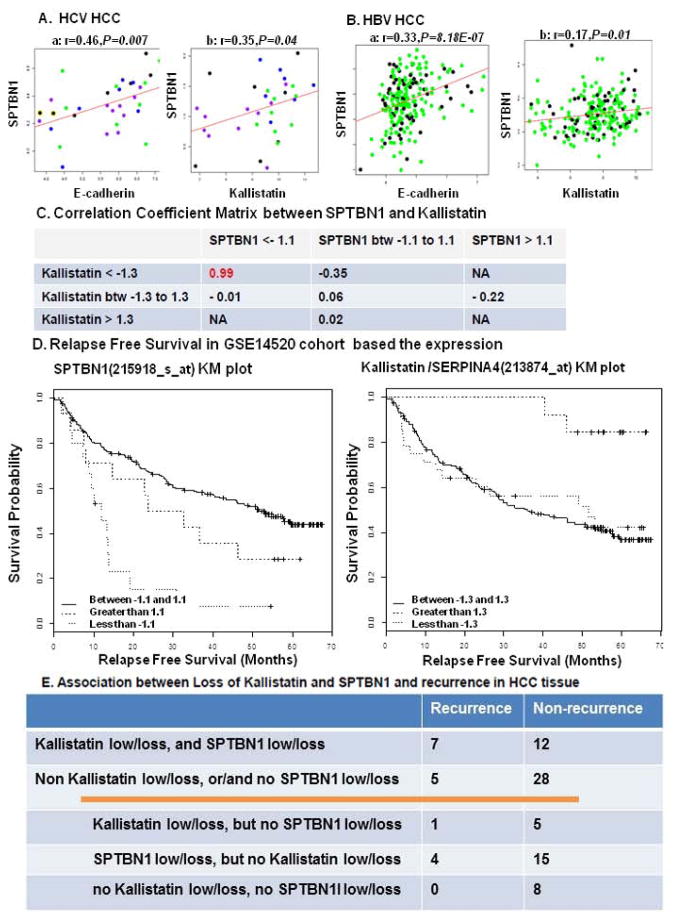
Figure 8.
The correlation of SPTBN1, E-cadherin and kallistatin was analyzed using published human gene array databases. A. GSE6764 correlation analysis was done on all 33 tumor samples from the Wurmbach study, including very early, early, advanced and very advanced HCC, which are shown as blue, green, black, and purple in the dot graph, respectively. B. GSE14520 study was done on 156 tumor samples with chronic carrier (CC) Hep B status and 56 tumor samples with active viral replication chronic carrier (AVR-CC) Hep B status, represented in green and black, respectively. The expression of SPTBN1 is positively and significantly correlated with E-cadherin and kallistatin in both HCV induced HCC and HBV induced HCC. “r” stands for correlation coefficient and “P” is for P value. B shows a positive correlation between decrease in kallistatin expression and decrease in SPTBN1 expression in HCC. We used the mean of the gene expression value as a reference to calculate fold change for each sample. We then separated the patients into sub-groups (low, medium and high expression) based on various-fold change cut offs ranging from a minimum-fold change value of 1 to maximum of 2. C and D demonstrate that loss of kallistatin or SPTBN1 suggests a decreased relapse free Survival (curve less than −1.3 in comparison to curve greater than 1.3 for kallistatin, curve less than −1.1 in comparison to curve 1.1 to −1.1 for SPTBN1). E shows the relationship between case number of recurrence and loss of SPTBN1 and Kallistatin protein evaluated by IHC stain in HCC patients after curative cancer resection.

