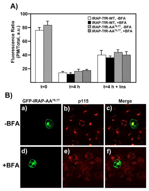
Fig. 3.
IRAP-TfR-WT and IRAP-TfR-AA76,77 both recycle from the cell surface back to an IRC that is BFA-insensitive. (A) Fully differentiated 3T3L1 adipocytes were transfected with 50 μg of either IRAP-TfR-WT or IRAP-TfR-AA76,77 and, 16 hours later, cells were stimulated with insulin and surface-labeled with the anti-TfR antibody on ice for 60 minutes. Cells were then washed with PBS and incubated at 37°C for 4 hours. For the BFA treatment, cells were incubated with BFA (5 μg/ml) during the last 30 minutes of the washout period and also during the second round of insulin treatment. Control cells were incubated with vehicle alone. Following the second round of treatment without or with insulin (100 nM, 30 minutes), cells were fixed, permeabilized and labeled with Texas-Red-conjugated secondary antibody, as described under Materials and Methods. The ratio of plasma membrane fluorescence:total fluorescence was determined using the Zeiss LSM software package (mean ± s.e.m. of three independent experiments). (B) Control experiments demonstrating the efficacy of 5 μg/ml BFA. Cells were transfected with 50 μg of GFP-IRAP-AA76,77 reporter construct and immediately plated in media either without (panels a-c) or with (panels d–f) 5 μg/ml BFA. After a 3-hour recovery period, cells were fixed and labeled with an anti-p115 monoclonal antibody, followed by Texas Red secondary antibody. a.u., arbitrary units.

