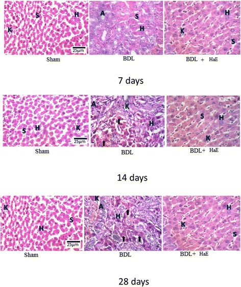
Figure 3.

Histological study of hematoxylin& eosin stained liver sections (400×) of Sham, BDL and Holothuria arenicola extract (HaE) treated rats. Sham treated liver showing normal hepatocytes (H) and separated by blood sinusoids (S). Bile duct ligated liver showing liver fibrosis indicated by adipocytes (A) and many collagen fibers (arrows). HaE- treated liver sections demonstrating the regeneration of liver parenchyma and light fibrosis.
