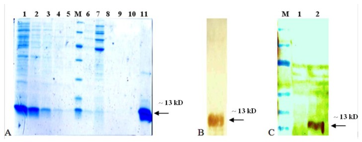
Figure 3.
Purification and identification of recombinant EDIII3. A SDS-PAGE analysis of purified EDIII3 protein. Different stages of purification on Ni-NTA affinity column; Lane1: cell lysate, lane 2-5: imidazole elution fractions, lane 6-10: washed fractions, lane 11: the pure EDIII protein after dialyzing against PBS buffer (before injection), lane M: Protein molecular weight markers (116, 66, 45, 35, 25, 18 and 14 kD). B Western blot analysis of purified EDIII3 which reacted with anti-His-Tag Mab. C Specific reactivity of recombinant EDIII3 with a mouse monoclonal anti-dengue antibody in western blotting analysis. Lane 1 is protein fraction from untransformed bacterial lysate (as negative control), lane 2 is protein fraction of EDIII3 expressing bacterial lysate, and lane M is protein molecular weight marker (175, 130, 95, 70, 62, 51, 42, 29, 22, 14 and 10 kD). The arrow indicates the position of the EDIII3 recombinant protein

