
Fig. 1.
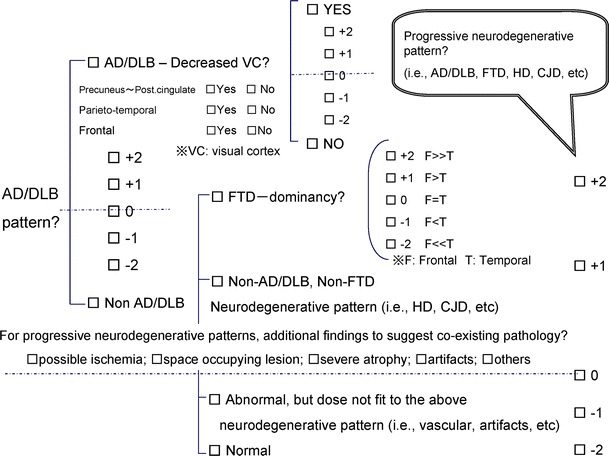
Check sheet used for the central image interpretation. Each expert was asked to report the findings on SPECT images and 3D-SSP Z score maps based on this diagnostic tree. First, the images were classified into AD/DLB pattern and non-AD/DLB pattern with confidence rating, and then sub-classified in each category. Additional findings to suggest co-existing pathology were also reported