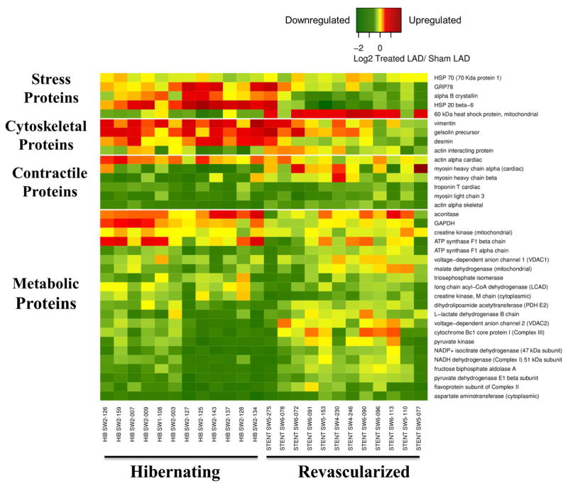
Figure 4. Heat Map Demonstrating Differential Expression of Proteins Correlating With Increased Flow and/or Function From Animals With Hibernating Myocardium.
Following revascularization and the alleviation of ischemia, up-regulated (red) stress and cytoskeletal proteins and glycolytic enzymes generally normalized (yellow). In contrast, many mitochondrial and contractile proteins remained depressed (green) 4 weeks after revascularization.

