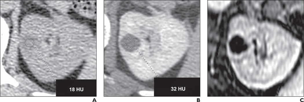
Fig. 3.
54-year-old woman with history of hepatitis C, cirrhosis, and 15-mm incidental left renal lesion.
A, Unenhanced CT shows attenuation of 18 HU.
B, Nephrographic phase CT shows attenuation of 32 HU (nephrographic minus unenhanced = 14 HU).
C, Fat-saturated dynamic contrast-enhanced T1-weighted gradient-echo MRI with subtraction shows absent internal enhancement, compatible with pseudoenhancement.

