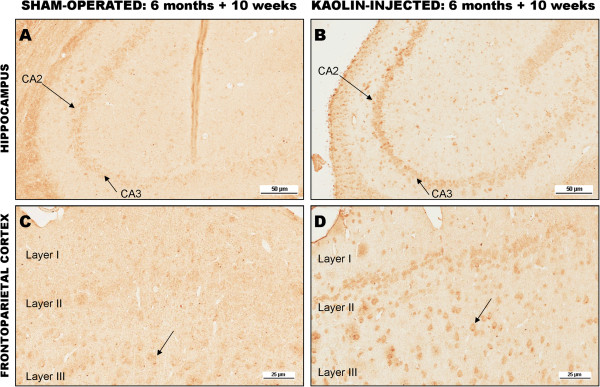
Figure 3.

Immunohistochemical staining for Aβ40. (A) Hippocampal neurons in a sham-operated six-month-old tgAPP21 rat (arrows). There is minimal immunoreactivity evident 10 weeks after sham-surgery, x80. (B) Hippocampal neurons in a hydrocephalic six-month-old tgAPP21 rat 10 weeks after kaolin injection demonstrating enhanced immunoreactivity in areas CA2 and CA3 (arrows), ×80. (C) Frontoparietal cortical neurons (arrow) in a sham-operated six-month-old tgAPP21 rat at 10 weeks post-surgery showing minimal Aβ40 immunoreactivity, ×200. (D) There is more robust neuronal immunoreactivity in the frontoparietal cortex in six-month-old tgAPP21 rats 10 weeks following kaolin injection (arrow), ×200.
