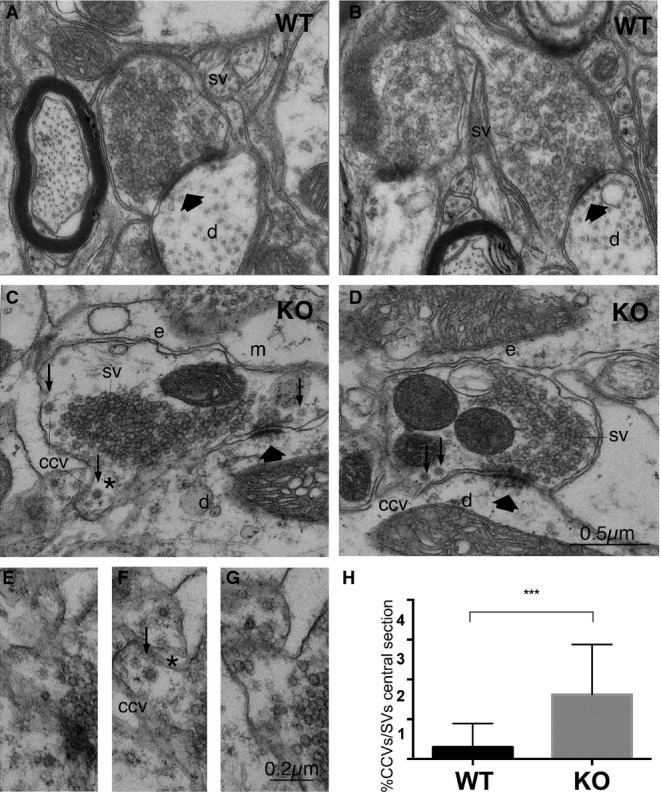
Figure 1.

- Electron micrographs of S-boutons establishing synapses on dendritic shafts in lamina IX of the mouse lumbar spinal cord of WT (A, B) or intersectin 1 KO mice (C, D). Scale bar: (A–D) 0.5 μm.
- Serial section from area marked in (C) (asterisk); CCV, free clathrin-coated vesicle. Scale bar: (E–G) 0.2 μm.
- Percentage of CCVs/total number of SVs (***P < 0.001; two-tailed unpaired t-test; n = 30, control and 32, intersectin 1 KO synapses from three mice of each genotype). Data are given as mean ± SD. SV, synaptic vesicles; d, dendritic shafts; e, endosome-like structures; m, mitochondrion; thick arrows indicate active zones; small arrows, CCVs.
