
Figure 1. In vitro and in vivo evidence that OSM binds to ECM.
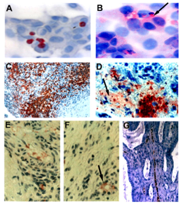
(A) OSM-expressing neutrophils were co-cultured with MDA-MB-231 breast cancer cells (10:1 ratio neutrophils to tumor cells), and no association of OSM with tumor cell ECM was apparent at 24 h. (B) Following 48 h incubation with neutrophils, OSM immobilized to the ECM of tumor cells was observed (arrow). (C–D) Low and high magnification photomicrographs of OSM expressing neutrophils in a colorectal tumor section. The colon cancer cells were negative for OSM expression, although a large population of OSM-expressing neutrophils that had infiltrated into the tumor was apparent (C). Upon high magnification, OSM associated with tumor ECM was observed (D; arrow). (E–F) OSM associated with the ECM of serial sections of an ovarian tumor. The similar patterns of OSM localized to the ECM of serial sections provided evidence against artifactual staining. (F) A circular orifice surrounded by OSM staining was apparent (arrow), which may represent a neutrophil canal. (G) An ovarian tumor section, which contains a neutrophil canal with OSM-expressing neutrophils.