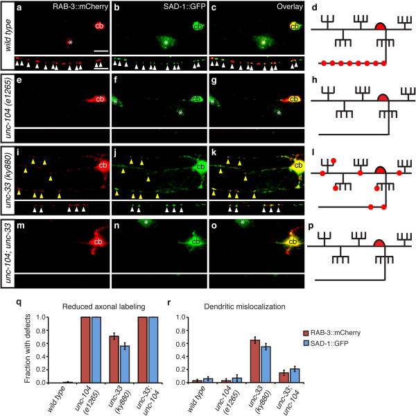
Figure 4. unc-104/KIF1A kinesin mislocalizes presynaptic proteins to dendrites in unc-33 mutants.
(a-p) Representative images of RAB-3::mCherry and SAD-1::GFP in PVD neurons of wild type (a-d), unc-104(e1265) (e-h), unc-33(ky880) (i-l), and unc-104 unc-33 (m-p) animals, with corresponding diagrams. For each set of fluorescence micrographs, the top panel is the maximum intensity projection of dendritic focal planes and the bottom panel is the maximum intensity projection of axonal focal planes. White and yellow arrowheads indicate axonal and dendritic puncta, respectively; ‘cb’ marks the PVD cell body, and asterisks mark gut autofluorescence. Anterior is at left and dorsal is up in all panels. Scale bar, 10 μm.
(q,r) Quantification of axonal localization defects (q) and dendritic mislocalization defects (r) of RAB-3::mCherry and SAD-1::GFP, as in Fig. 1; n>30 animals/genotype. Error bars indicate s.e.p.

