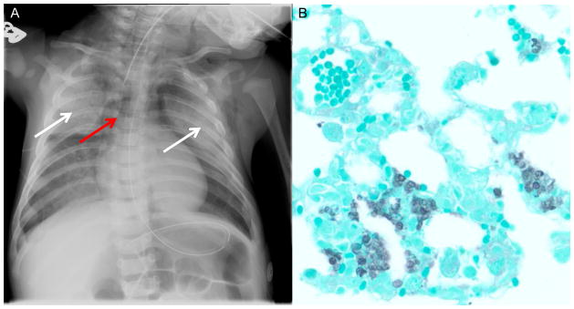
Figure 1.

Radiographic and microbiologic diagnosis of Pneumocystis. A. Chest radiograph of child with X-linked severe combined immunodeficiency showing bilateral ground glass infiltrates (white arrows) and air bronchograms consistent with Pneumocystis pneumonia. Note that this patient also has a pneumomediastinum (red arrow) with air dissecting into the soft tissue of the neck and an absent thymic shadow in the mediastinum consistent with athymia. B. Gomori-methenamine silver stain (GMS) on Pneumocystis infected mouse lung, showing lung architecture (green) with Pneumocystis organisms (black) filling the alveolar spaces.
