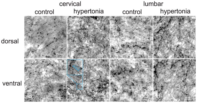
Figure 5.

Serotonergic fibers in dorsal (first row) and ventral (second row) horns in spinal cord of control (A,C,E,G) and hypertonic kits (B,D,F,H). Density of serotonergic fibers was visibly higher in hypertonic group. Course of serotonergic fibers could be reconstructed following the beaded lines on anti-serotonin immunostaining. Objective 40x, scale bar 25 μm.
