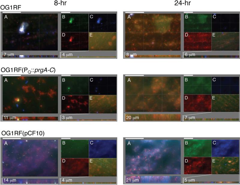
FIG. 6. The Prg proteins and eDNA mediate early biofilm development.
Macroscopic images of sections of biofilms produced by strains inoculated on PMMA coupons at the onset of cCF10 pheromone induction and incubated for 8 h (left panels) and 24 h (right panels) in TSB medium. Intact cells were stained with hexidium iodide (HI, red), eDNA and lysed cells were stained with GelGreen (green), and EPS was stained with calcofluor white (blue). Panels A-E are identified for the 8 h biofilm images of strain OG1RF and similarly-arranged for the other strains and time points analyzed. Panels A: Three color-merged images; scale bars, 50 μm. Below: Sagittal projections showing biofilm thicknesses with measured values shown in μM; scale bars, 3.125 μm. Panels B-D: Matched sets to biofilms in panel A showing separate staining of (B) GelGreen, (C) HI, (D) calcofluor white. Panels E: Biofilms were treated with DNase at the onset of pheromone induction, stained with GelGreen, HI, and calcofluor white, and the three-color merged images are presented, with biofilm thicknesses presented below with scale bars equivalent to those used in Panel A. Strains: OG1RF, (OG1RF(pCF10), OG1RF(pINY1801) which expresses PQ::prgA-C.

