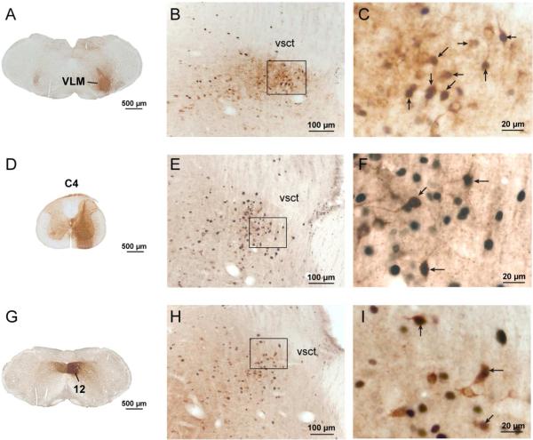
Figure 7.
A series of photomicrographs illustrating the projections of the Kolliker-Fuse nucleus to respiratory-related sites. Panels A-C show c-Fos-activation with CO2 exposure in the projection to the ventrolateral medulla; D-F show projections to the phrenic motor level of the spinal cord; and G-I demonstrate projections to the hypoglossal motor nucleus. Injection sites into the brain (A, G) or spinal cord (D) of animals exposed to CO2 are shown on the left. The middle panels show a low magnification photomicrograph through the KF nucleus (B, E, H), and a higher magnification image on the right (C, F, I) showing the area in the box in B, E, H from a hypercapnic animal in each series. Note the large percentage of doubly-labeled neurons (indicated by arrows, brown cytoplasm demonstrating CTb, black nucleus stained for c-Fos) in each case. Few if any doubly labeled neurons were seen in control brains (Figs 6, 8, 9). Abbreviations: C4, fourth cervical spinal segment; VLM: ventrolateral medulla; 12 hypoglossal motor nucleus; vsct: ventral spinocerebellar tract.

