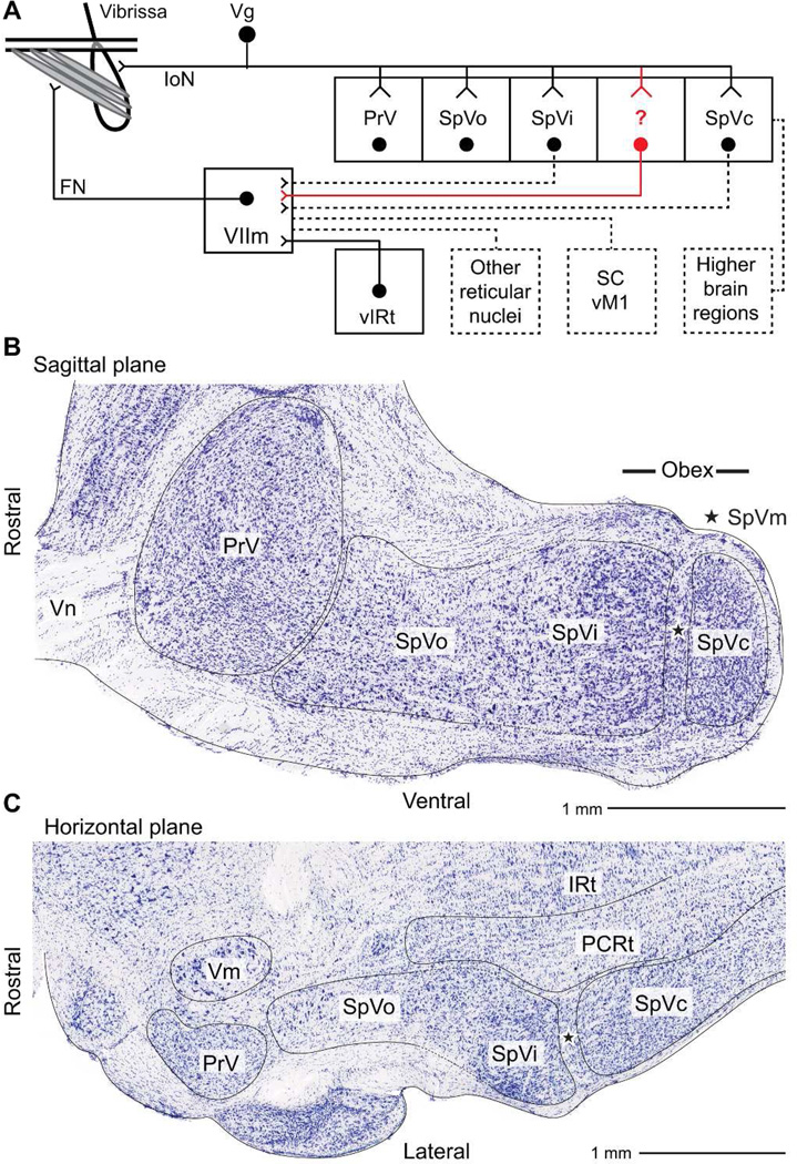
Figure 1. Organization of the trigeminal nuclei.
A: Schematic of trigeminal brainstem nuclei. Sensory afferent neurons transmit signals from the mystacial pad via the infraorbital nerve (IoN) of the maxillary branch of the trigeminal nerve (Vn), through the trigeminal ganglion (Vg), and terminate throughout the trigeminal nuclear complex, which includes the principal trigeminal nucleus (PrV), and the spinal trigeminal nuclei oralis (SpVo), interpolaris (SpVi), and caudalis (SpVc). A fifth nucleus, spinal trigeminal nuclei muralis (SpVm) and its connections are the subject of this report (red-colored). Neurons in the trigeminal nuclear complex project within trigeminal brainstem, to facial motor nucleus (VIIm), and to higher brain areas in thalamus, cerebellum, and superior colliculus. VIIm motoneurons also receive input from a vibrissa pattern generator in the vibrissa zone of the intermediate reticular formation (vIRt) and from disparate cortical and subcortical nuclei. VIIm motoneurons project to extrinsic and intrinsic musculature of the face for vibrissa motor control. B: Nuclear outlines, based on classical cytological differences, overlaying Nissl-stained sections in the sagittal and horizontal planes. The distinct gross morphology of each of the trigeminal nuclei and several neighboring nuclei are outlined. PCRt refers to the parvocellular region of the reticular formation, IRt to the intermediate region of the reticular formation, and Vm to the trigeminal motor nucleus. The star (⋆) highlights the region that contains SpVm.

