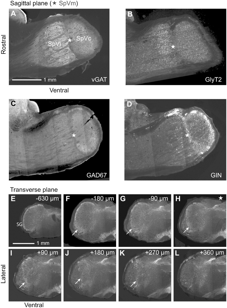
Figure 2. Sagittal and transverse views of inhibitory neurons within and near SpVm.
A–D: Sagittal sections from transgenic animals that express EGFP driven by particualr promoters: VGAT is the vesicular Gaba transporter, GlyT2 is the glycine transporter, GAD67 is the glutamic acid decarboxylase, and GIN is a line with eGFP-expressing inhibitory neurons. The star (⋆) highlights the region that contains SpVm. E: Transverse sections from GIN animals; SG is substantia gelatinosa and the arrows point to SpVm. Distances are from the location of SpVm in panel F with positive numbers in the rostral direction.

