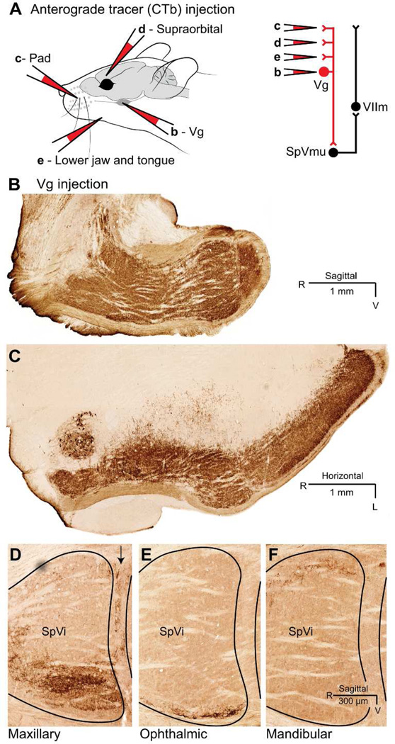
Figure 3. Sensory afferent axons terminate in the spinal trigeminal nuclei.
A: Schematic of the location of cholera toxin B (CTb) injections. The letter above each injection pipette indicates the panel it represents. Red-colored connections in the circuit diagram indicate the projections under examination. Injections were performed in independent animals. B–C: Representative sagittal (panel B) and horizontal (panel C) sections of trigeminal brainstem after bolus injections of CTb into Vg. Diaminobenzidine reaction product (dark brown) indicates axons and terminals, which occur throughout the trigeminal nuclear complex, Vm, and PCRt. D–E: Central afferent axonal terminations following focal injections of CTb in the regions of face innervated by the three trigeminal nerve branches: maxillary (panel D), ophthalmic (panel E), and mandibular (panel F), The border between nuclei SpVi and SpVc shows arborization after mystacial pad injection only, which labels the maxillary branch exclusively. Outlines show SpVi and the rostral edge of SpVc.

