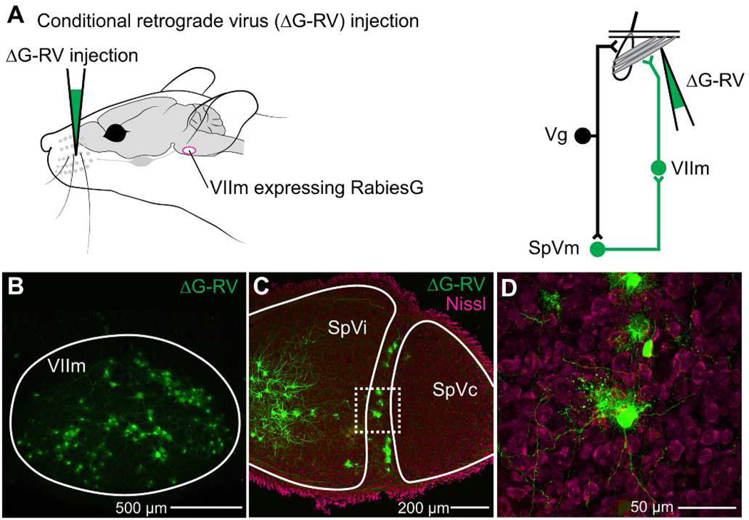
Figure 5. Retrograde labeling of SpVm by modified Rabies virus.
A: Injection strategy for premotor neuron labeling. ΔG-RV was pressure injected into the facial musculature of juvenile RΦGT mice and is transported transynaptically to facial motoneurons. Rabies glycoprotein is only expressed in the presence of cholineacetyltransferase, which is present in VIIm motoneurons using the Cre-Lox system. This permits active rabies to be recapituated in labeled facial motoneurons for subsequent labeling of premotoneurons. Schematic of vibrissa and the circuit under investigation are used throughout. B: Rabies-labeled (green) motoneurons are robustly labeled in lateral VIIm (sagittal section). C: Monosynaptically-connected premotor neurons in nuclei SpVo, SpVi, and SpVm, labeled by ΔG-RV (green) and counterstained with a fluorescent Nissl (Neurotrace red). D: Magnified images of a set of SpVm neurons (box in panel C), showing dendritic arborization and strict alignment along the border of SpVi and SpVc.

