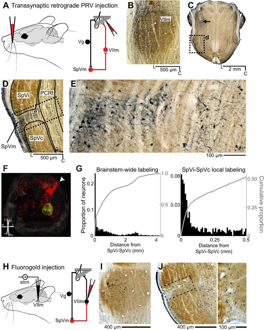
Figure 6. Retrograde labeling of SpVm by pseudorabies virus and FluoroGold.
A: Injection strategy. Pseudorabies virus (PRV) was injected into the intrinsic and extrinsic musculature of the left mystacial pad. B: Positive labeling in lateral VIIm ipsilateral to injection after 48 hours, horizontal slice. Large motoneurons are robustly labeled, as are parts of superior salivatory nucleus, rostral to VIIm. Medial divisions of VIIm are unlabeled. C: Representative brainstem slice showing PRV-positive neuron labeling. The most robust and densest labeling is in ipsilateral SpVm (box). PRV labeling is also visible in the genu of VII at this level, dorsal to VIIm (arrow). D: Magnification of box in c. Ipsilateral nuclei SpVm and PCRt are robustly labeled. E: Magnification of box in Panel D. VIIm-projecting SpVm neurons (left) are smaller and have more confined dendrites than the Golgi-like labeled neurons in PCRt (right). F: Volumetric reconstruction of brainstem from aligned, serial sections (see methods). Red dots indicate locations of 2251 PRV-positive, non-VIIm neurons, of which 29% (646 of 2251) are in ipsilateral SpVm (white arrowhead). VIIm (outlined in yellow) contains primary labeled motoneurons whose individual cell bodies are not shown for clarity; brainstem is outlined in gray. G: Distribution of PRV-positive neurons relative to the centerline between SpVi and SpVc (arrow in panel F). The top distribution has a bin size of 50 µm while the bottom has a bin size of 10 µm. H: FluoroGold injection strategy. After targeting lateral VIIm by eliciting vibrissa movement in response to microstimulation, tracer was iontophoresed. I: FluoroGold labeling in VIIm motoneurons. Lateral VIIm is strongly labeled, while medial VIIm labeling is absent. J: FluoroGold labeling of nuclei SpVm, PCRt, and IRt. The magnified and rotated image (box) shows neurons along the border between SpVi and SpVc.

