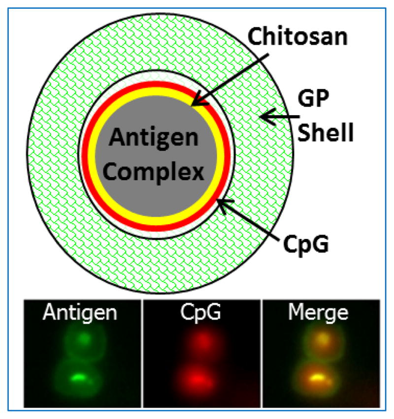
Figure 2. Glucan particles (GPs).

Top: Schematic showing the “layer-by-layer” design of a GP containing complexed antigen and CpG-rich DNA. Bottom: Epifluorescent photomicrographs of two abutting GPs loaded with antigen (fluorescently labeled green with FITC) and CpG (fluorescently labeled red with TRITC).
