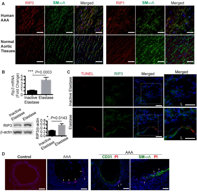
Figure 1. Increased receptor interacting protein 3 (RIP3) expression and cell necrosis in abdominal aortic aneurysm tissues.
(A) Representative photographs of human aneurysmal and normal aortic cross sections that were double stained for RIP3 (or RIP1, red) and smooth muscle α-Actin (SM-αA, green). n=4. Scale bars=50μm. (B) Real-time PCR (n=8) and Western blotting (n=4) analyses of Rip3 expression in C57BL/6 mouse arteries perfused with elastase or heat-inactivated elastase on Day 14 post-perfusion. Data are mean±SEM. (C) Representative photographs of aortic cross-sections harvested from C57BL/6 mice on Day 3 post-surgery. Sections were co-stained for RIP3 and TUNEL. Higher magnified views of highlighted regions were shown on the right. L indicates lumen. n=3. Scale bars=50μm. (D) Representative photographs of aortic cross sections of normal or elastase-perfused aortae (Day 7 post-perfusion). Propidium iodide (PI) was administered to mice via intraperitoneal injection 2 hours prior to sacrifice. Following PI staining, aortic sections were also stained for CD31 or SM-αA. n=3. Scale bars=100μm.

