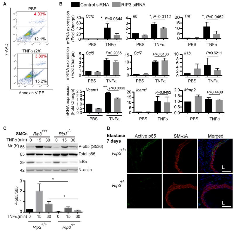
Figure 5. RIP3 is involved in TNFα-induced cytokine expression in aortic SMCs.
(A) Aortic SMCs isolated from C57BL/6 mice were treated with TNFα (10ng/ml) for 2 h. The absence of cell death induced by TNFα at this concentration was confirmed by flow cytometry. n=2. (B) Aortic SMCs were transfected with control or RIP3-specific siRNA. After 48 hours, cells were stimulated with 10ng/ml TNFα for 2 hours for cytokines and Mmp2 or 2ng/ml TNFα for 4 hours for Vcam1 and Icam1. mRNA levels were determined by Real-time PCR. Data are mean±SEM. n=4. (C) Aortic SMCs isolated form Rip3+/+ and Rip3-/- mice were treated with 10ng/mL TNFα for indicated time. Cell lysates were subjected to Western blot analysis with indicated antibodies. *P<0.05. Data represent mean±SEM. n=4. (D) Representative photographs of aortic cross-sections harvested from mice on Day 7 post-perfusion with elastase. Sections were co-stained for active p65 and SM-αA. L indicates lumen. n=3. Scale bars=50μm.

