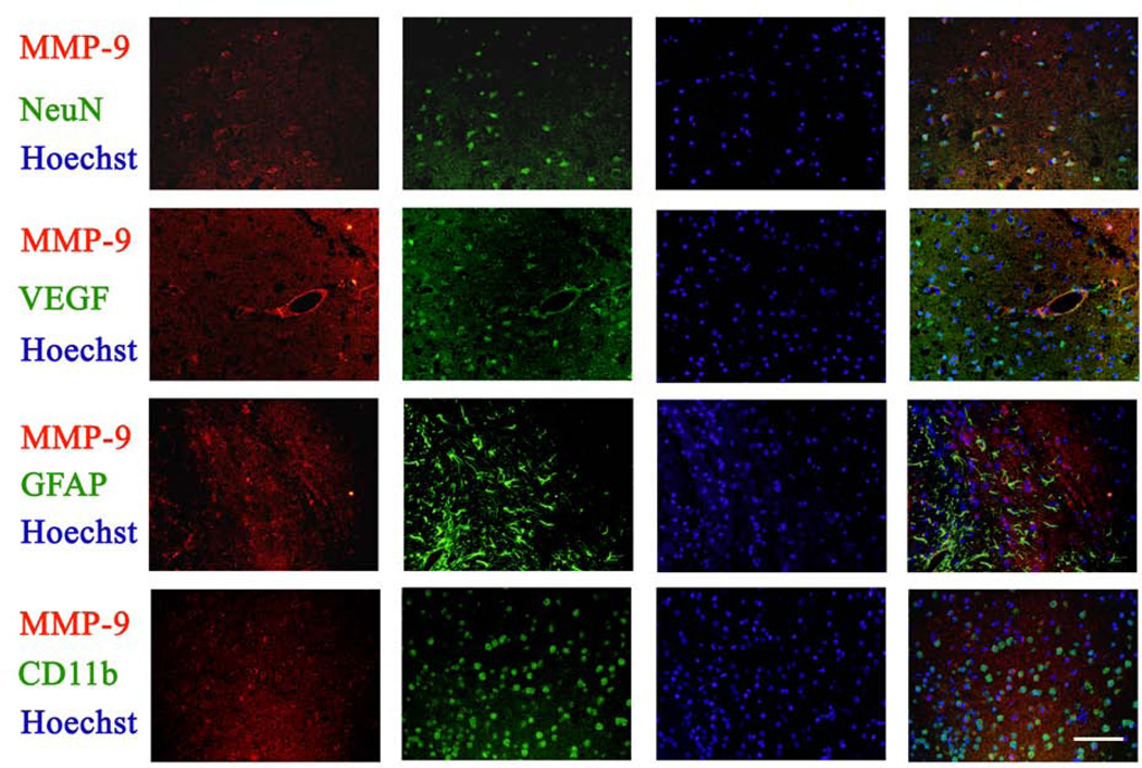
Fig. 5. Expression of MMP-9 in the brain tissues.
Coronal sections at Bregma −0.58 mm were obtained from animals subjected to brain ischemia only for immunofluorescent staining of MMP-9 (red), NeuN (green), VEGF (green), GFAP (green) and CD-11b (green). Images were taken in the ischemic Fr1 areas. The merged panels also include Hoechst staining (blue) to show cell nuclei. Bar = 100 µm.

