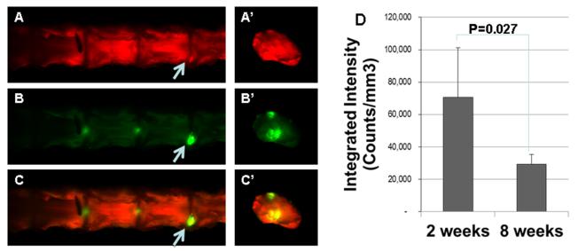
Figure1. Allogeneic rabbit articular chondrocytes survived in the degenerating rabbit disc.
Chondrocytes labeled with infrared dye were injected into the degenerating disc. Left Panels: the rabbit spines and discs were imaged at 2 weeks post injection. A & A’: coronal view of the spine and transverse view of an individual disc showing tissue contour; B & B’: infrared dye labeled cells; C & C’: overlay of the above panels. Right Panel: intensity of the infrared dye at 2 and 8 weeks post injection.

